
Article
Preparation and Characterization of DPPDA-Eu3+ Doped Silica Fluorescent Nanoparticles
Dongguang Yin *, Li Zhang, Chunjuan Xie, Binhu Liu, Le Zhang
College of Environmental and Chemical Engineering, Shanghai University, Shanghai, 200444.
* Corresponding author. Email: ydg@shu.edu.cn
Citation: D. Yin, et al., Preparation and Characterization of DPPDA-Eu3+ Doped Silica Fluorescent Nanoparticles. Nano Biomed. Eng., 2010, 2(1): 40-44.
DOI: 10.5101/nbe.v2i1.p40-44.
Abstract
A new ligand of 4,7-diphenyl-1,10-phenanthroline-2,9-dicarboxylic acid (DPPDA) and Eu3+chelate compound of this ligand were prepared. Then DPPDA-Eu3+ doped silica fluorescent nanoparticles of DPPDA-Eu3+/SiO2 with primary amino groups on their surface were developed using a water-in-oil (W/O) microemulsion technique. Characterizations by transmission electron microscopy, fluorescent spectra, fluorescent molecules leaking experiments and photostable experiments show that the nanoparticles are spherical, monodisperse, and uniform in size (80±8 nm in diameter). The nanoparticles have high fluorescent signal and high photostability. When the nanoparticles were dispersed in aqueous solution with continuously ultrasound for 4 h, no obvious leakage of fluorescent molecules were observed. As a novel fluorescent probe, the nanoparticles are expected to be applied in highly sensitive bioassays systems such as time-resolved fluorescence immunoassay, biosensor and biochip.
Keywords: Europium chelate; 4,7-Diphenyl-1,10-phenanthroline-2,9-dicarboxylic acid; Silica nanoparticles
1. Introduction
Recently, application of nanoparticles in bioassay has been rapidly developed [1-3]. Several sorts of nanoparticles, such as gold nanoparticles [4-5], semi-conductor nanoparticles for example, quantum dots [6-8], magnetic nanoparticles [9-11], carbon nanotubes [12-16], and luminophore-doped silica nanoparticles have been developed and widely applied in various bioassay systems. Compared to traditional organic fluorescent dyes, nanometer-sized luminescent probes have advantages of highly fluorescent signal, excellent photostability and biocompatibility. Based on big Stokes displacement and long lifetime of fluorescence signal, time-resolved fluorescence analysis(TRFA) is one of the most sensitive bioassay techniques. Using lanthanide chelate-doped silica nano-particles as luminescent probe is very favorable for TRFA due to their good water-solubility, biocompati-bility, easy preparation and surface modification [17-18].
2. Materials and Methods
2.1 Materials and instruments
Bathocuproine(98 %), N-chlorosuccinimide, Eu2O3, triton X-100, 3-aminopropyl(trimethoxyl) silane (APT-MS), tetraethyl orthosilicate (TEOS), n-hexanol, cyclohexane and ammonium hydroxide (28-30 wt %) were purchased from Sigma. All of these regents were analytical grade. The 1H NMR spectra were recorded on a Bruker AVANCE 500 spectrometer (Switzerland). UV-vis absorption spectra were measured on a Hitachi 3010 UV-vis spectrophotometer (Japan). The transmission electron microscopy (TEM) was measured on JEOL-200CX transmission electron microscope(Japan). TRFIA was measured on a Thermo Varioskan Flash Multifunction Microplate Reader(USA).
2.2 Preparation of DPPDA
0.10 g bathocuproine, 0.25 g N-chlorosuccin- imide, 0.50 mg benzoyl peroxide in 2.5 mL carbon tetra-chloride were mixed, refluxed for 6 h with stirring, then cooled to room temperature and filtered . After rotary evaporating, residue was dissolved in 2.5 mL chloroform, washed with 2.5 mL Na2CO3 saturated aqueous solution, then organic layer was collected. After crude product was dried over night with anhydrous MgSO4, evaporated and dried, desired product of 2.9-bis (trichloromethyl) - 4.7 – diphenyl -1.10-phenanthroline was obtained(0.12 g, 75.7 % yield). 0.088 g 2.9-Bis(trichloromethyl) - 4.7 – diphenyl-1.10-phenanthroline and 0.39 mL sulfuric acid were mixed, heated with stirring for 2 h at 80 C. After cooling with ice, 0.19 mL ice water was added and the mixture was heated with stirring for 1 h at 80 C. When the reaction mixture was added to 6 mL ice water, a shallow yellow precipitate was generated and it was collected. After filtrating, water washing and vacuum drying, the final product of DPPDA was obtained(0.062 g, 94.2 % yield). Anal. Calcd for C26H16N2O4 (DPPDA): C 74.28, H 3.84, N 6.66; found C 74.35, H 3.76, N 6.52. 1HNMR ((CD3)2SO, 500MHz) δ: 7.618(m,10H, benzene-H), 8.027(s,2H,benzene-H), 8.285(s,2H, pyridine-H), 3.6(s, 2H,COOH), 2.5(s, DMSO).
2.3 Preparation of DPPDA-Eu3+chelate and DPPDA-Eu3+/SiO2 nanoparticles
1.1 mg Eu2O3 was dissolved in 0.5 mL of 6 M HCl and heated until a white crystal was generated. Then the crystal was dissolved in 0.5 mL anhydrous ethanol. When 5.0 mg DPPDA was dissolved in 0.5 mL anhydrous ethanol, 0.5 mL of 6.25×10⁻⁵ M EuCl3 in anhydrous ethanol was added drop by drop with stirring. The reaction was allowed to continue for 10 h at room temperature. After filtrating and vacuum drying at 37 ℃for 1 h , the desired product of DPPDA-Eu3+ was obtained (2.8 mg, 46.7 % yield). Anal. calcd for (C26H16N2O4)2Eu·15H2O(DPPDA-Eu3+): C 49.45, H 4.913, N 4.437, O 29.16; found C 49.63, H 4.902 , N 4.310, O 29.23. 3.0 mg of DPPDA-Eu3+, 100 μL of TEOS, and 0.55 mL of water were added to a 50 mL round-bottom flask with stirring. Then a water-in-oil (W/O) microemulsion containing 2.24 mL of Triton X-100, 2.23 mL of hexanol, 9.32 mL of cyclohexane was added to the flask with vigorous stirring. Finally, 5 μL of APTMS and 100 μL of NH4OH(28 %) were added to the mixture. The reaction was allowed to continue for 24 h. The pure DPPDA-Eu3+/SiO2 nanoparticles were obtained after isolating by adding acetone to break the microemulsion , centrifuging , ultrasonically washing three times with ethanol and water respectively, and vacuum drying at 37℃ for 1 h. Pure silica nano-particles without primary amino groups on their surface were also prepared by the method of water-in-oil (W/O)micro-emulsion.
2.4 Phobleaching experiments
To evaluate photostability of the nanoparticles, photobleaching experiments of the nanoparticles and pure DPPDA-Eu3+ chelate compound were performed in 0.05 M Tris-HCl buffer( pH7.8) using a 100 W xenon lamp as an excitation source. Fluorescent intensities were recorded at every 10 min interval for a period of 40 min.
2.5 Fluorescent molecules leaking experiments
3.0 mg of the nanoparticles were dissolved in 10 mL water and the mixture was dispersed by ultrasound continuously. At every 1 h, 1 mL of the suspending solution was taken out and centrifugated. After cen-trifugal separation, the precipitate was redissolved in 1 mL water, ultrasonic dispersed, and then the fluo-rescence intensities were measured on a Hitachi F-7000 spectrophotometer.
3. Results and Discussion
The structures of DPPDA and DPPDA-Eu3+ chelate compound were shown below.

3.1 TEM image of the nanoparticles
The TEM image of the DPPDA-Eu3+/SiO2 nano-particles was shown in Figure 1. The nanoparticles are spherical and uniform in size (80±8 nm in diameter) with excellent monodisperse.
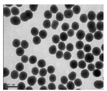
Figure 1. TEM image of the nanoparticles

Figure 2. Excitation and emission spectra of DPPDA-Eu3+/SiO2 nanoparticles A and a,0.3 mg/mL)and DPPDA-Eu3+ chelate(B and b, 3.3 ug/mL) in a 0.05 mol/L Tris-HCl buffer( pH 7.8).(A, B: excitation spectra; a, b: emission spectra)
3.2 Fluorescence spectroscopy of the nanoparticles
As shown in Figure 2, fluorescence spectra of the DPPDA-Eu3+chelate and the DPPDA-Eu3+/SiO2 nano-particles display a similar profile in a 0.05 M Tris-HCl buffer (pH 7.8). All of them show the same excitation and emission maximum wavelengths at 300 and 615 nm, respectively. The emission patterns of pure DPPDA-Eu3+ chelate and the nanoparticles are typical for the Eu3+ fluorescent compounds, and three sharp emission peaks at 596, 615, and 694 nm correspond to the 5D0 →7F1,2,4 transitions of Eu3+. As can be seen from the spectra, the DPPDA-Eu3+chelate and the DPPDA-Eu3+/SiO2 nanoparticles all have a wide exci-ation wavelength and a sharp emission peaks with 10 ~ 15 nm of half-peak width, and all have a 300 nm of large stokes displacement which is favorable to effec-tively eliminate short-lived scattering light and back-ground noises.
3.3 Photostability of the nanoparticles
As shown in Figure 3, results of photobleaching experiments revealed that the fluorescent intensity of the DPPDA-Eu3+chelate was decreased approximately 22.3 % after 40 min of continuous excitation, whereas the fluorescent intensity of the nanoparticles was only decreased 0.9 %. The high photostability of the nano- particles is caused by the fact that the DPPDA-Eu3+ chelate in the nanoparticles is coated surroundingly by silica which isolates the chelate from the outside envir-onment such as solvent molecules and free radicals caused by light exposure and, therefore, effectively protects the chelate from photodecomposition.
3.4 Fluorescent molecules leaking experiments of the nanoparticles
As shown in Figure 4, the fluorescent intensity of the nanoparticles was only decreased approximately 1.4 % after continuously ultrasonic 1 h in aqueous solution and, with the extension of time, there are no obvious fluorescent intensity decreasing was observed. These results indicate the nanoparticles are stable in aqueous solution.
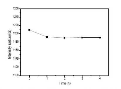
Figure 3. Photostability curve of the DPPDA-Eu3+/SiO2 nanoparticles(A) and DPPDA-Eu3+ chelate(B)

Figure 4. Fluorescent molecules leaking curve of the nanoparticles

Figure 5. UV-vis absorption spectra of APTMS(A), nanoparticles of DPPDA-Eu3+/SiO2 (B), pure silica nanoparticles without amino groups on their surface (C)
3.5 Confirmation of amino groups on surface of the nanoparticles
Based on the fact that a blue-violet compound was generated which has an absorption peak at 570 nm when hydrated ninhydrin reacted with amino com-pound [19], confirmation of amino groups on surface of the nanoparticles was investigated. As shown in Figure 5, APTMS and the nanoparticles all have a obviously absorption peak at 570 nm after they reacted with hydrated Ninhydrin, but the pure silica nanoparticles without amino groups on their surface has non-absorption at 570 nm. Results demonstrate that amino groups had been directly introduced to the surface of the nanoparticles through a copolymerization reaction between APTMS and TEOS. Since these active amino groups are introduced to the surface of the nanoparticles during the preparation process, the nanoparticles can be directly used to conjugate with biological molecules with no need of complicated surface modification.
4 Conclusions
DPPDA-Eu3+/SiO2 fluorescent nanoparticles having amino groups on their surface were prepared using a water-in-oil (W/O) microemulsion technique, and characterized with TEM, spectroscopy, photobleaching experiments and fluorescent molecules leaking experiments. Results show that the nanoparticles are spherical and uniform in size(80±8 nm in diameter), monodiesperse, high fluorescent intensity, high photo-stability, and high stability in aqueous solution. The nanoparticles, as a new capable fluorescence probe, are expected to be applied in various highly sensitive biological detection systems such as time-resolved fluorescence immunoassay, biochips and biosensors.
Acknowledgments
Financial support from the Shanghai Pujing Program, Shanghai Nano Program (No. 0752nm024), and Shang-hai Leading Academic Discipline Project (No. S30109) is gratefully acknowledged.
Rererences
Received 26 January, 2010; accepted 18 February, 2010; published online 5 March, 2010.
Copyright: (c) 2010 D. Yin et al. This is an open-access article distributed under the terms of the Creative Commons Attribution License, which permits unrestricted use, distribution, and reproduction in any medium, provided the original author and source are credited.

|
Nano Biomedicine and Engineering. Copyright © Shanghai Jiao Tong University Press |
 |