
Article
The Pharmacokinetic Profile of Freeze-dried Cyclosporine A-Eudragit S100 Nanoparticle Formulation in Dogs
Yang Liu 1, Zhigang Fang1, Yagen Chen 1, Jiankang Zhang 1, Xuenong Zhang1,*, Qiang Hong2
1College of Pharmaceutical Science, Soochow University, Suzhou, 215123, China
2Department of Pharmaceutics , School of Pharmaceutical Science , Peking University , Beijing 100083, People’s Republic of China
* Corresponding author: zhangxuenong@163.com (Xuenong Zhang)
The pharmacokinetic profile of freeze-dried cyclosporine A-Eudragit S100 nanoparticles (CyA-S100-NP)was studied with a random two-way crossover study in dogs. The drug blood concentration was determined by internal standard HPLC method after oral administration of CyA-S100- NP and Neoral. Pharmacokinetics parameters were calculated by 3P97 program. The concentration-time data were fitted as a two-compartment open model. The AUC of CyA-S100-NP was higher than that of Neoral(P<0.05), while the CL significantly decreased(P<0.05). The relative bioavailability of CyA-S100-NP were 135.9% compared with Neoral. The bioavailability of CyA was significantly improved. CyA-S100-NP was a potential drug for developing a new CyA nanoparticles solid formulation.
Keywords: Cyclosporine A, Nanoparticle, Pharmacokinetics, Bioavailability, HPLC
Citation: Y. Liu et al, The pharmacokinetic profile of freeze-dried cyclosporine A-Eudragit S100 nanoparticle formulation in dogs, Nano Biomed. Eng. 2011, 3(1), 53-56.
DOI: 10.5101/nbe.v3i1.p53-56.
Introduction
Cyclosporine A (CyA), an immunosuppressant, has been used for preventing organ transplant rejection in humans and the treatment of systemic and local autoimmune disorders. However, it is known that the oral bioavailability of CyA is usually very low with a high inter- and intra-patient variability due to the poor absorption, which is related to the relatively high molecular weight, very high lipophilicity and extensive metabolism. Moreover, the major limitation of CyA remains hepatotoxicity, nephrotoxicity, central nervous system toxicity and gastroenteric reaction (Cohen et al.,1984; Thomson et al., 1991; Tjai et al., 1991).Most studies have been performed to decrease the above mentioned side effects and increase the therapeutic efficacy of CyA. CyA-nanoparticles have been used extensively as a formulation for improving the bioavailability of poorly water-soluble drugs due to its small particle size and unique uptake mechanisms. In addition, smaller particles are more readily absorbed (Hillyer et al., 2001). By virtue of enterically soluble polymers (Eudragits® S100 ), which can dissolve at specific pH values, cyclosporin A pH sensitive nanoparticles could target different parts of the gastrointestinal tract and protect metabolism of the incorporated drug, meanwhile, not containing Cremophor RH40. Cremophor RH40 exert nephrotoxic and may cause anaphylactoid reactions (Luke et al., 1987). It has been reported that pH-sensitive nanoparticles improved the absorption and bioavailability of CyA. (Dai et al., 2004; Wang et al., 2004).Yang et al. demonstrated CyA- Eudragit S100-Nanoparticles significantly improved the bioavailability of CyA after oral administration to rats (YANG et al., 2008). The pharmacokinetics of CyA have been investigated extensively in the dogs (Ford et al., 1999; Lee et al., 2001; El-Shabouri et al., 2002), as this species has been used as the model for organ transplantation in humans. Moreover, the pharmacokinetics and metabolic patten are similar in dogs and humans (Venkataramanan et al., 1988; Vickers et al., 1992). The bioavailability and pharmacokinetics of CyA from these nanoparticles in comparison with the commercial microemulsion formulation (Sandimmune® Neoral) were assessed in dogs.
2. Methods
2.1. Preparation of CyA-S100-NP capsules
An h y d r o us e t h a n o l c o n t a i n i n g C y A/ E u d r a g i t ® S100 (1:4, w/w) were injected as soon as possible into stirring water containing 125 mg Poloxamer 188 at room temperature (organic phase/aqueous phase, 2:5 ). The mixture was stirred at 400 rpm, 10 min later, ethanol residues were left to evaporate in a 60˚C water bath for 3 h and then CyA-S100-NP colloids were obtained. The CyA- NP was concentrated to 8 mg CyA per milliliter before applied. Aliquots (3 mL) were placed in sealed vials in the presence of 2.5% lactose and frozen at -70˚Cfor 24 h. Then, the samples were immediately placed on the shelves of the freeze- drying chamber (ALPHA 1-4/LD, Marin Christ Co., Germany). Sublimation lasted 28 h without heating. Finally, CyA-S100-NP capsules were obtained (ZHANG et al., 2008).
2.2. Particle Size Analysis
CyA-S100-NP were diluted 10-fold by ultrapure water, prior to examination. The analysis of particle size was performed by dynamic light scattering (Malvern Instruments Ltd, UK) at the wavelength of 670 nm and the temperature of 25˚C.
2.3. Drug Loading and Encapsulation Efficiency
Prepared suspension of CyA-NP was filtered through a 0.45 μm filter to remove insoluble polymer residues and CyA microcrystals. Then, 8 ml of the filtered suspension was ultracentrifuged at 250, 000 × g for 60 min under 10˚C and the supernatant was sampled (YANG et al., 2008). CyA content in the filtered suspension and in the supernatant were analyzed by HPLC. The HPLC system is composed of two pumps (LC-20AD, Shimadzu, Japan), UV-vis detector set at 210 nm. The chromatographic column used was BDS HYPERSIL C18 (5 μm in 4.6 mm×250 mm, Thermo Fisher Scientific, USA) maintained at 70˚C. The mobile phase consisted of acetonitrile/ (methanol: water) {55/45 (1:1) ,v/v} and the flow rate was 1.3 ml/min-1. The injection volume was 20 ml. The yield, encapsulation efficiency (EP%) and drug loading efficiency (LD%) were calculated.
2.4. Sample Preparation
CyA in blood was determined by a reversed phase HPLC method, as mentioned above. Briefly, 0.5 ml of blood sample was added to a centrifuge tube, then 50 μl of internal standard (cyclosporine D, CyD) in methanol at a concentration of 30 μg•mL-1 and 1 ml of hydrochloric acid (180 mmol•L-1) were added. After vortex mixing for 1 min, 5 ml of ether was added. The extraction was carried out by shaking the tube horizontally for 15 min at 100 rpm, centrifuging it for 15 min at 4000 rpm, and separating the ether phase to another centrifuge tube. After 1 ml of sodium hydroxide solution (95 mmol•L-1) and 2.5 ml of 1% sodium pyrosulfite solution were added, the extraction was repeated by the same procedure as described above. The ether layer was transferred into a clean tube and evaporated to dryness under nitrogen at 40˚C. The residue was reconstituted with 150 μl of acetonitrile/water (70/30, v/v). The reconstituted blood samples were washed three times by 1 ml of n-hexane (LI et al., 1999). 20 μL of residual solution was injected into the HPLC system for the determination of CyA and CyD.
2.5. Method validation
50 μL CyA methanol solution with concentrations of 0.5025,1.005,5.025,10.05,20.1,30.15 μg•mL-1 was added to 0.5 ml blank whole blood giving final concentrations of 0.05025,0.1005,0.5025,1.005,2.01,3.015 μg•mL-1, in the presence of internal standard CyD (30 μg•mL-1). Each blood standard was then treated with the same preparation procedure described above. Quantitation was done by determination of peak-area ratio of CyA/CyD (A/Ai) against the drug concentrations. The concentrations of unknown samples were determined by using the linear regression line of the concentration of the calibration standard versus peak-area ratios.50 μL CyA methanol solution with concentrations of 0.5025,1.005,2.01 μg•mL-1 was added to 0.5 ml blank whole blood, then each blood standard was taken through the sample preparation procedure described above. 20 μL residual solution was injected into the HPLC system to calculate the recovery of CyA. Five times one day, and repeated for 5 days to estimate intra-day and inter-day precision.
2.6. Experimental Design
Six healthy dogs were randomly divided into three groups. CyA-S100-NP and Neoral (equivalent to 10 mg•kg-1of CyA) were orally administered to dogs in each group, respectively. The dogs were fasted overnight prior to the experiments and continued for further 4 h after administration, but allowed free access to water. The washout period was two weeks. At predetermined time intervals (0.5, 1, 1.5, 2, 4, 6, 8, 12, 24 and 36 h), blood samples (1 ml) were drawn from the antecubital vein into heparinized tubes and stored in the frozen state for subsequent analysis.
2.7. Statistical Analysis
Pharmacokinetic parameters were estimated using 3P97 (a computer program produced by the Committee of Mathematical Pharmacology of the Chinese Society of Pharmacology). The maximal concentration (Cmax) and the time to maximal concentration (t max ) were obtained directly by observation. The total area under the concentration-time curve (AUC) from time zero to infinity was calculated by the trapezoidal rule method. The statistical and graphical analyses were accomplished using commonly available commercial software packages. Differences between the mean values were analyzed by multiple comparison test where P<0.05 was considered to be significance. All results were expressed as mean±standard deviation.
3. Results
3.1 Characterization of CyA-S100-NP
The mean particle size of CyA-S100-NP was 52.7±4.6nm. CyA-S100-NP had a loading efficiency of 22.9±0.2% and displayed high encapsulation efficiency over 99.5%. The yield was 98.1±0.2%.

Fig. 1. HPLC chromatograms of CyA in blood. A blank blood;B blank blood containing CyA and CyD D;C blood sample;1 CyA;2 CyD
Under the described chromatographic conditions, CyD can be separated from CyA. As Fig 1 shown, There was no endogenous substance in the blood which would interfere with the determination of CyA and CyD, and there was no impurity introduced in the sample preparation procedure. The linearity of CyA was determined in the range 0.05025-3.015 µg•mL-1 with a regression coefficient of 0.9994. The regression equation of concentration of CyA in the blood: C = 2.5098Ai-0.1044.
Table 1. The recovery and precision of CyA in blood ( x ±S)
|
Added concentration (μg•mL-1) |
Recovery (%) |
RSD (%) |
||
|
Extract |
Method |
Intra-day |
Inter-day |
|
|
0.5025 |
94.92±8.522 |
102.7±6.267 |
6.103 |
9.083 |
|
1.005 |
104.9±8.402 |
94.12±1.942 |
2.063 |
4.544 |
|
2.010 |
109.1±2.868 |
95.43±2.575 |
2.698 |
4.640 |
Table 2. Pharmacokinetic parameters of CyA after a single oral dose of Neoral and CyA-S100-NP ( x ±S)
|
Parameters |
Neoral |
CyA-S100-NP |
|
AUC(μg•mL-1•h) 0-36h |
10.66 ±2.01 |
14.49 ±3.00* |
|
AUC(μg•mL-1•h) 0-∞h |
12.77 ±3.08 |
17.08 ±2.66* |
|
Cmax (μg•mL-1) |
1.08 ±0.07 |
1.38 ±0.22* |
|
Tmax (h) |
2.17 ±0.93 |
2.25 ±0.88 |
|
MRT (h) |
10.94 ±0.94 |
11.16 ±0.97 |
|
V/F(c) (L•kg-1) |
4.76 ±0.98 |
5.40 ±0.47 |
|
CL(L•h-1) |
0.80 ±0.20 |
0.57 ±0.09* |
|
Fr (%) |
—— |
135.9 |
Relative bioavailability:Fr % = (AUCtest×DNeoral /AUCNeoral×Dtest)×100% * P﹤0.05 vs. Neoral
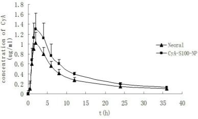
Fig. 2. The mean blood concentration-time curve of CyA following oral administration of CyA-S100-NP and Neoral to dogs (n=6)
The mean extract recovery rate of CyA was over 94% and the method recovery rate was 94.1%-102.7%, the intra-day and inter-day precision were less than 10% (Table 1). While the recovery rate of CyD was 96.58±6.563%.
The blood concentration profile of CyA in dogs after oral administration of a 10mg•kg-1 dose is presented in Fig.2. A two-compartment open model was adequate to describe the blood pharmacokinetics of CyA in dogs. The data obtained from the individual dogs were analysed by the two compartment model, using 3P97 software for final parameter estimates. Corresponding pharmacokinetic parameters are listed in Table 2. The results obtained revealed that the Cmax and AUC of CyA from CyA-S100-NP show significant difference (P<0.05) when compared with those of Neoral. The relative bioavailability CyA-S100-NP to the reference was 135.9%. In addition, the CL was decreased significantly (P<0.05). There were no significant differences in other pharmacokinetic parameters of CyA between two formulations.
Methodological approaches to determining the concentration of CyA in whole blood include high performance liquid chromatography (HPLC) and a variety of immunoassay-based methods, such as radioactive immunoassay (RIA), fluorescence polarization immunoassay (FPIA). Although immunoassay-based methods are simple to use, the cross-reactivity of the antibody with the metabolites leads to overestimation of CyA concentrations in blood by these methods. The HPLC method described here is specific, sensitive, reproducible and is applicable to pharmacokinetic studies of CyA. The treatment of blood samples of CyA include liquid- liquid extraction and solid-phase extraction method. The liquid-liquid extraction method is accurate and reproducible, although complicated. In this paper, the treatment of whole blood samples was established based on the study of LI et al. (LI et al., 1999), including acidification, extraction, alkalization, washing and other steps. During the experiment, the amout of N-hexane and frequency of washing played a key role in removal of impurity. Under chromatographic conditions mentioned above, CyD could be separated from CyA. The results showed that accuracy and recovery were consistent with the determination of biological samples. This method is applicable to analysis of CyA concentration in dogs, and also to pharmacokinetic and bioavailability study in other species. The pharmacokinetics of CyA-S100-NP in dogs shows, the bioavailability of CyA was significantly improved. The Cmax and AUC from CyA-S100-NP were significantly increased as comparision of those from Neoral, while CL decreased significantly (P<0.05). The release profiles of cyclosporin A pH sensitive nanoparticles exhibit significant pH-sensitivity, there was a burst release of CyA from nanoparticles at specific part of the gastrointestinal tract (YANG et al., 2008), which is possible to decrease metabolism of the drug by gastrointestinal enzymes, increasing the oral bioavailability of CyA. Due to not only high oral bioavailability, but also long term stability, CyA-S100-NP could be a more effective nanoparticle solid formulation for oral delivery of poorly water-soluble CyA.
This work was supported by the National Key Technology R&D Program in the 11th Five Year Plan of China (2006BAI09B00), National Basic Research Program of China (973 Program No. 2007CB935800), and National Key Program of New Drug innovation (No. 2009zx09310-001).
1. Cohen, D.J., Loertscher, R., Rubin, M.F. 1984. Cyclosporine A: a new immunosuppressive agent for organ transplantation. Ann. Intern. Med. 101: 667–682.
2. Dai Jundong, Nagai Tsuneji, Wang Xueqing, et al. 2004. pH- sensitive nanoparticles for improving the oral bioavailability of cyclosporine A. Int. J. Pharm. 280: 229-240. doi:10.1016/ j.ijpharm.2004.05.006
3. El-Shabouri M.H., 2002. Positively charged nanoparticles for improving the oral bioavailability of cyclosporin-A. Int. J. Pharm. 249: 101-108. doi:10.1016/S0378-5173(02)00461-1
4. Ford J., Woolfe J., Florence A.T., 1999. Nanospheres of cyclosporin A: poor oral absorption in dogs. Int. J. Pharm. 183: 3-6. doi:10.1016/ S0378-5173(99)00049-6
5. Hillyer J.F., Albrecht R.M., 2001. Gastrointestinal persorption and tissue distribution of differently sized colloidal gold nanoparticles, J. Pharm. Sci. 90 (12): 1927–1936. doi:10.1002/jps.1143
6. Lee Eun- Jin, Lee Sa- Won, Choi Han- Gon, et al. 2001. Bioavailability of cyclosporin A dispersed in sodium lauryl sulfate- dextrin based solid microspheres. Int. J. Pharm. 218: 125-131. doi:10.1016/S0378-5173(01)00621-4
7. LI HS, YE GQ, ZHAO JL, et. al. 1999. Improved high performance liquid chromatographic determination of cyclosporine A in whole blood samples of rat and rabbit. West China J Pharm Sci. 14(2): 89- 92.
8. Luke DR, Kasiske BL, Matzke GR, et al. 1987. Effect of cyclosporin on the isolated perfused rat kidney[J]. Transplantation 43(6): 795- 799.
9. Thomson, Neild, Cyclosporin: use outside transplantation. Brit. Med. J., 1991, 302: 4–5. doi:10.1136/bmj.302.6767.4
10. Tjai J.F., Webber I.R., Back D.J. 1991. Cyclosporin metabolism by the gastrointestinal mucosa[J]. Br. J. Clin. Pharmacol. 31(3):344- 346.
11. Venkataramanan, R., Wang, C.P., Habucky, K., et al. 1988. Species specific cyclosporine metabolism. Transplantation proceedings 20: 680–683.
12. Vickers, A.E., Fischer, V., Connors, S., et al. 1992. Cyclosporine A metabolism in human liver, kidney and intestine slices – comparison to rat and dog slices and human cell lines. Drug Metabolism and Disposition 120: 802–809.
13. Wang Xue-qing, Dai Jun-dong, Chen Zhen, et al. 2004. Bioavailability and pharmacokinetics of cyclosporine A-loaded pH- sensitive nanoparticles for oral administration. J. Control. Release. 97: 421-429.
14. YANG Zhi-qiang, Pan Ping, 2008. Xu Jie, et al. Study on Preparation of CyA-Eudragit S100-Nanoparticles and Its Characteristics in vivo and in vitro. Chin Pharm J. 43(23):1798-1803.
15. YANG Zhi-qiang, Xu Jie, Pan Ping, et al. 2008. Study on preparation and releasing characteristics of Cyclosporin A pH sensitive nanoparticles in vitro. Chinese Journal of New Drugs. 17(20): 1784- 1786.
16. ZHANG Xue-nong,YANG Zhi-qiang, Pan Ping. A self-emulsifying formulation of freeze-dried cyclosporine A nanoparticles and its preparation method. China Patent,200710191876.2008-06-11.
Copyright:(c) 2011 Y. Liu, et al. This is an open-access article distributed under the terms of the Creative Commons Attribution License, which permits unrestricted use, distribution, and reproduction in any medium, provided the original author and source are credited.

|
Nano Biomedicine and Engineering. Copyright © Shanghai Jiao Tong University Press |
 |