Research Article
Muhammad Aqeel Aslam 1, 2, Muhammad Asif Munir 3, Rauf Ahmad 3, Muhammad Samiullah 3, Nasir Mahmood Hassan 4, Shahzadi Mahnoor 5, Daxiang Cui 1, 6
1 Institute of Nano Biomedicine and Engineering, Shanghai Engineering Research Center for Intelligent Instrument for Diagnosis and Therapy, Department of Instrument Science & Engineering, School of Electronic Information and Electrical Engineering, Yantai Information Technology Research Institute of Shanghai Jiao Tong University, Shanghai Jiao Tong University, 800 Dongchuan Road, Shanghai 200240, P.R. China.
2 Electrical Engineering Department, GIFT University,Gujranwala, Pakistan.
3 Electrical Enngineering Department, Swedish College of Engineering & Technology, Rahim Yar Khan, Pakistan.
4 Electrical Engineering Department, Khawaja Fareed University of Engineering & Information Technology, Rahim Yar Khan, Pakistan.
5 Information and Commmunciation Engineering Department, UESTC, Chengdu, China.
6 National Engineering Research Center for Nanotechnology, 28 Jiangchuan Road, Shanghai 200241, P.R. China.
* Corresponding author. E-mail: dxcui@sjtu.edu.cn
Received: Oct. 18, 2021; Accepted: May 8, 2022; Published: May 11, 2022
Citation: Muhammad Aqeel Aslam, Muhammad Asif Munir, Rauf Ahmad, Muhammad Samiullah, Nasir Mahmood Hassan, Shahzadi Mahnoor, and Daxiang Cui, Deep Neural Networks for Prediction of Cardiovascualr Diseases. Nano Biomed. Eng., 2022, 14(1): 81-89.
DOI: 10.5101/nbe.v14i1.p81-89.
Abstract
In recent years, a huge extent of data that contains hidden information is collected by the health care industries. Deep Neural Networks (DNN) have been employed to obtain appropriate decisions and effective results. The obtained results have been validated using confusion matrix and region of interest. In this work, we have used fourteen parameters for the prediction of cardiovascular disease (CVD) of 303 volunteers. The proposed predictive technique predicts that the chance for prediction of the risk level of cardiovascular disease. In this work, the prediction method using deep neural networks showed the highest accuracy. Our proposed method has outperformed the existing methods and can be combined with multimedia technology.
Keywords: Artificial intelligence, Deep learning, Heart disease prediction, Neural network
Introduction
According to the World Health Organization (WHO), Cardiovascular disease (CVD) is the leading cause of death globally. Around 18 million people die due to CVD in one year. CVDs are related to disorder of the heart and blood vessels. There are few other factors which are also responsible for the heart disease (HD), these factors include lack of physical activity, smoking, obesity and irregular eating times [1-3]. CVDs are further divided into cerebrovascular disease, coronary heart disease, rheumatic heart disease and other conditions. Heart attacks and strokes caused the 80% of the deaths associated to CVDs [4]. Moreover, the young people are also suffering from CVDs, as more than one-third deaths are caused due to a kind of heart disease. In 19th and 20th century, the physicians used to diagnose the working of the heart by listening the heartbeat directly from the chest, which is not only a very nonscientific way but also an unethical approach [5]. The instrument named stethoscope was first developed in 1816, and is commonly used medical instrument to diagnose the diseases, which includes the chest infection [6]. This device allows the subject to measure the heart rate in real time. The heart varies in the male and female gender, and in newly born babies the heart rate is 120 bpm at the rest position [7]. A clear pattern is produced by the normal heart beat. Whereas, the heart beat produced by the heart patients has a noisy pattern, which indicates the heart is not working properly. The physicians practice in the routine to make the correct diagnosis. However, it is difficult to diagnose the heart disease for non-clinical person and inexperience even by the use of stethoscope. To diagnose the hart disease accurately, several years of experience and a long term practice is needed, which is relatively hard to obtain [8]. Once a person is suffering from the heart disease, it may not be able to receive proper oxygen and nutrients, which is required for the heart muscles to function at their best. This will limit the flow of oxygen to the heart [9]. With passage of time, the arteries become thin and blood can’t flow smoothly. This may produce the heart attack due to the blockage of blood [10]. Like other cure disease, if the heart disease is not diagnosed in the initial stages, the patients may suffer. Heart transplant and stent surgery are commonly used in advanced treatments. These methods widens the arteries and blood start flowing smoothly [11]. This treatment can reduce the chance of the stroke and heart attack. There are few clinical methods, which are mostly deployed to diagnose the heart diseases. These methods include Chest X-Ray, Blood tests, Electrocardiography (ECG), and Electrocardiogram (EEG) [12]. CVD is difficult to diagnose at the early stage due to it’s vague symptoms. Moreover, most of the heart patients diagnosed in the advanced stage of the disease. However, if the CVD is diagnosed at the early stages, the effect of the disease can be minimal by convenient medications and proper exercise to make sure that the blood is circulating properly. Therefore, it is very important to diagnose the CVD at the initial stage for successful, treatment. Computer Aided Technologies for the diagnosis of fatal diseases have received significant attention from the researchers [13]. Machine learning (ML) has laid down the foundation of artificial intelligence (AI). AI has become the essential tool for the medical researchers. ML comprises of three classes, which are supervised ML techniques, unsupervised ML techniques, and semi-supervised ML techniques. Supervised ML methods are commonly used to predict the disease. In general, supervised algorithms are used when the class labels are known, whereas, the unsupervised algorithms are used for regression problems. Semi-supervised ML technique is latest addition in the Artificial Intelligence (AI) branch, it is the combination of the supervised ML techniques and unsupervised ML techniques. Data mining techniques discovers the hidden pattern from the data input, this data input is non-linear separable. A few studies have already been carried out by the researchers using data mining approaches for disease prediction [14-16] To diagnose the disease, Artificial Neural Network (ANN) [17], Naive Bayes Classifier (NBC) [18], Decision Tree [19], K-Nearest Neighbor (K-NN) [20], Support Vector Machine (SVM) [21] and Random Forest (RF) [22] are commonly used tools for supervised ML methods. In general, the use of AI in the medical field has increased the diagnostic accuracy 15% to 20% [23]. Deep Learning (DL) is the most recent and advance tool of ML technique [24]. In last few years, the DL has gained a tremendous attention from the scientist and computer engineers to overcome the health related problems. DL algorithms are frequently used in computer vision, pattern recognition, and classification tasks. DL is an emerging tool which is used for disease diagnosis in the medical field [25]. In 2018, HassanT.T et al., developed a hardware based system on Field Programmable Gate Array using Numeral Virtual Generalizing Random Access Memory (NVG-RAM) to diagnose heart disease. They achieved 88% for multiclass heart diseases [26]. In 2020, Muhammad Yar et al., developed an early and accurate detection and diagnosis method using intelligent computational model, and achieved an accuracy of 95.8% for ANN based prediction system using three hidden layers [27]. In 2012, Andullah et al., proposed a data mining method that can be used to predict the coronary heart disease. They developed neural network using RF classifier. They achieved 63.33% accuracy [28]. In 2010, Srinivas et al. developed a neural network using NBC to predict the heart disease [29]. In 2013, Nahar et al. presented a medical driven approach for the prediction of heart disease [30]. They used different data mining techniques. This research indicates Medical Feature Selection (MFS) is promising technique for heart disease diagnostics. In 2013, Chaurasia et al. demonstrate data mining techniques for the early prediction of heart disease [31]. In 2009, Das et al. developed neural network ensembles to diagnose the heart disease effectively [32]. In 2021, Yazdani, et al., proposed a novel approach for heart disease prediction using strength scores with significant predictors. They achieved confidence score of 98% in predicting the disease [33]. However, they reduced their study to 8 significant features. They also have not mentioned the selectivity, sensitivity, specificity and other parameters, which may be very vital in any deep neural network based architecture. In 2020, Almustafa et al., proposed a method to predict the heart disease and classifiers sensitivity analysis. They achieved an accuracy of 93. 6585% using Decision Tree (DT) based architecture [34].However,they have used only four atributrs out of thirteen. In 2020, Shah et al., proposed machine learning techniques to predict the heart disease. The authors used forteen attributes to visulaize the performance of the different algorithms..They achieved an overall accuracy of 88.175% for NBC based architecture and 90.789% for K-NN based architectures [35]. In 2021, Jindal, et al., developed machine learning based architectures to predict the heart disease. They developed K-NN, RF Classiifier and Logistic Regressson based architectures to predict the heart disease. They achieved an accuracy of 88.5% with K-NN and Logistic Regression [36]. The list of abbreviation is listed below in the Table 1. The proposed model is a combination of unsupervised and supervised techniques. It grouped the featured data suing unsupervised method and developed a model that can predict the outcome by suing supervised learning method. The formation of the rest of the paper is as: Materials and Methods are discussed in section 2. This part comprises of Dataset, Deep Sparse Autoencoder (DSAE), Performance Evaluation, and Experimental Setup. Results and Discussions are discussed in section 3. Conclusion about this study is done in section 4.
Table 1 Enumeration of the symbols used in this paper
|
Symbol
|
Descripton |
Symbol |
Description |
|
AI |
Artificial Intelligence |
ANN |
Artificial Neural Network |
|
CVD |
Cardiovascular Disease |
DNN |
Deep Neural Network |
|
DT
|
Decision Tree |
DSAE |
Deep Stacked Sparse Autoencoder |
|
EEG
|
Electrocardiogram |
HD |
Heart Disease |
|
MFS
|
Medical Feature Selection |
ML |
Machine Learning |
|
RF
|
Random Forest |
ROC Curve |
Receiver Operating Characteristic Curve |
|
SVM
|
Sipport Vector Machine |
WHO |
World Health Organization |
|
AUC
|
Area Under Curve |
K-NN |
K-Nearest Neigbor |
|
DL
|
Deep Learning |
NBC |
Naïve Bayes Classiifer |
|
ECG
|
Electrocardiography |
SAE |
Sparse Autoencoder |
Experimental
Dataset
In this work, the dataset was obtained from the UCI Irvine Machine Learning repository. Black et al., [37] has provided the information in detail about the dataset in 2013. The dataset contains 303 clinical samples, who volunteer participated in this study. There were more than seventy attributes but 14 attributes in each sample were used for this study. We excluded three attributes, which were patient identification number, age social security number, as all these three attributes are not useful and easily be excluded from the dataset. Only 11 attributes were used to analyze the performance of the proposed Deep Neural Network. The CVD prediction system is a binary classification, which contains two output, either the person is suffering from the CVD or the person belongs to the non-CVD class. 165 persons were affected by the CVD, whereas, 138 people belong to the healthy class.
Deep sparse autoencoder
Deep Learning has become a very popular tool in diagnosis of fatal diseases. The sparse autoencoder is an emerging tool in artificial intelligence to sole the medical related applications. The autoencoder comprises of two parts; namely encoder and decoder. It uses unsupervised learning technique to extract the hidden features from the input data using feed-forward neural network. In deep structure, the autoencoder has played an ultimate role [38-42]. The simple architecture of sparse autoencoder has been shown in Fig. 1. Like other neural networks, Sparse Autoencoder (SAE) also consists of three layers: namely input layer followed by hidden layer and L2 the output layer. In this study, we have used two hidden layers, and this model is called deep sparse autoencoder. The cost function of the SAE comprises of three terms as shown in Equation 1.
![]()
In Equation 1,![]() is the constant for L2 weight Regularization and
is the constant for L2 weight Regularization and ![]() is the constant for the sparsity Regularization. The L2 weight Regularization term is calculated as follows:
is the constant for the sparsity Regularization. The L2 weight Regularization term is calculated as follows:
![]() . (2)
. (2)
Equation 3 represents the sparsity Regularization term.
![]() (3)
(3)
The proposed architecture of Deep Sparse Autoencoder (DSAE) complete training in two phases, in first phase the unsupervised greedy layer-wise initialization mad in second phase fine tuning is carried out using supervised learning schemes. The first SAE tries to minimize the reconstruction error and second SAE use the output of the first SAE as an input with the hidden activation. In last, these deep autoencoder connected to the Softmax later for the final fine tuning phase [43]. The flowchart of the proposed DSAE algorithm is shown in Fig. 2.
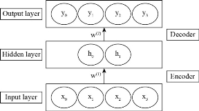
Fig. 1 Basic structure of autoencoder, it contains two parts, encoder and decoder at output layer we reconstruct the input data by minimizing the x and y whereas hidden layer is represented by h. w(1) and w(2) are the weight values
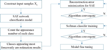
Fig. 2 Flow chart of the proposed DSAE algorithm for cardiovascualr disease prediction.
Performance evaluation
The dataset was divided into two parts, training part consist of 70% and test part consist of 30%. We performed some preprocessing steps to make sure that each attribute has a same influence on the performance of the proposed classifier. After the preprocessing steps, the data was fed as an input to the classifier. There are several parameters that can be used to evaluate the performance of the develop neural network. In this study, we have used confusion matrix and receive operating characteristics curve to measure the accuracy of the proposed architecture. A confusion matrix indicates the actual and predicted classification for each class. We can easily visualize from the confusion matrix that how many samples are misclassified in each class. As already discussed, this is a binary classification problem, the confusion matrix composed of four possible outcomes:
(TN): Those instances where the model has accurately predicted that person is healthy;
(TP): Those instances where model has accurately diagnosed the heart disease;
(FN): Those instances where model has misclassified the patients and recognized them as healthy;
(FP): Those instances where the model has misclassified the healthy persons into patients.
The following parameters were used to evaluate the performance of the proposed classifier.
![]() . (4)
. (4)
![]() . (5)
. (5)
![]() . (6)
. (6)
![]() . (7)
. (7)
![]() . (8)
. (8)
The Receiver Operating Characteristic (ROC) curve was also used to observe the performance of the proposed classifier. This curve is plotted between TP rate and FP rate for each threshold. The more the area under the curve, the higher will be the performs of the classifier. For good accuracy, the Area Under Curve (AUC) has to be close to one, if this value is close to zero this indicate that the classifier needs to be redesigned by using different values of the parameters, for 100% accuracy, the AUC will be equal to 1.
Table 2 Parameters used in this study
|
First sparse autoencoder |
||||
|
Number of neurons |
Sparsity regularization |
L2 weight regularization |
Decoder function |
Sparsity proportion |
|
50
|
8 |
0.0001 |
Pure line |
0.25 |
|
Second sparse autoencoder
|
||||
|
Number of neurons
|
Sparsity regularization
|
L2 weight regularization |
Decoder function |
Sparsity proportion |
|
30
|
8 |
0.0001 |
Pure line |
0.05 |
Experimental setup
For this work, we wrote the routines for the deep sparse autoencoder neural network using MATLAB 2017b. Our developed neural network contains two hidden layers each hidden layer has different number of neurons. The detailed information of the developed neural network is shown in the Table 2. The repeatability and reliability are the important factor of any neural network, to make sure the accurate diagnosis, we performed the experiment 30 times and the results are the average of these thirty experiments. All the parameters were kept constant during these thirty experiments.
Results and Discussion
The deep learning algorithm shows good performance even on the modest dataset. Our developed model has assured the freedom of the training samples from the testing samples set. The dataset contains 303 samples, we used 258 samples for training + validation and 45 samples were used to evaluate the performance of the classifier. The test dataset contains 45 samples. We compared our proposed method developed by Deep Neural Networks with Shallow Feedforward Artificial Neural Network to confirm the effectiveness of the developed method. Fig. 2, represents the confusion matrix of the training data, validation data, test data and complete dataset for single layer ANN based CVD prediction system. First we select the parameter for the ANN. The parameters include number of hidden layers and number of neurons present in the hidden layer. We train the ANN using single layer and once the training has completed, we tested the test data to evaluate the performance of the ANN based CVD prediction system. The ANN based classifier produces an accuracy of 84.5% for the complete dataset and 85.9% for training dataset, 82.2% for the test data. The ANN based predictive system has predicted 256 samples correctly, however, the model has misclassified 47 samples. The performance of the ANN based model depends upon the architecture as well as the parameters, number of hidden layers, number of neurons are key in the ANN. However, for deep ANN results may vary due to the number of layers. Fig. 4 shows the performance graph of the training data, validation data, and test data of the ANN based architecture. The best validation was achieved at 14 epochs, and the best validation performance was 0.17264. Fig. 5, represents the ROC curves for the ANN based classifier. Four different ROC curves were drawn using TPR and FPR, each for training data, validation data and test data. The fourth ROC curve is the combination of first three curves. In second experiment, we develop a multi-layer feedforward neural network using scaled conjugate gradient method. We used two hidden layers, the first hidden layer contains 20 number of neurons and second layer consist of 10 number of neurons. This architecture performed better than the single layered neural network. Fig. 6 represents the confusion matrix for the two layered architecture. This architecture yields an overall accuracy of 85.8%. This method predicted 150 and 110 samples of patients and healthy persons, respectively. This method misclassified 15 and 28 samples from each class. After completing the training process, the model produces an accuracy of 83.6% for the test data. Fig. 7 shows the ROC curve for the multi-layer feedforward neural network. We can clearly visualize that the AUC for the multi-layer architecture based neural network is more than the single layered neural network architecture. In the third experiment, we developed deep sparse autoencoder based neural network to distinguish the heart disease patients from the healthy persons. We tried different architectures for the deep sparse autoencoder to develop a reliable and fast algorithm for the prediction of heart disease. Fig. 8 shows the results for deep sparse autoencoder based neural network in the form of confusion matrix. We trained the neural network first using deep sparse autoencoder, once the network was trained we tested the developed neural network with different dataset (training data and test data are different). Each autoencoder was trained for 1000 epochs. The deep sparse autoencoder produced an overall accuracy of 94.5%, this model predicted 35 heart disease samples correctly, and 51 samples of healthy persons were accurately classified. During the test phase, 3 and 2 samples were misclassified from patient’s and healthy person’s class, respectively. Table 3 summarizes the results of different experiments conducted during this study and also compares the results aceived in this study with the previous published results. From the above table we can visualize that the sparse autoencoder has outperformed the shallow neural network based model in terms of performance. However, the deep sparse autoencoder require more time to complete the training, once the training is completed, the results of the deep neural network is better than the other state of art technologies.
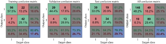
Fig. 3 Confusion matrices using artificial neural network with 1-layer for heart disease prediction.
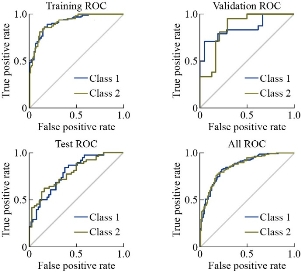
Fig. 4 Performance curve of training data, validation data.
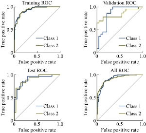
Fig. 5 Receiver operating characteristics curve using artificial neural network with 1-layer for heart disease prediction.
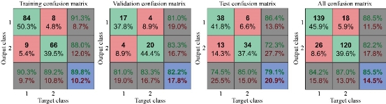
Fig. 6 Confusion matrices using artificial neural network with multi-layer for heart disease prediction.
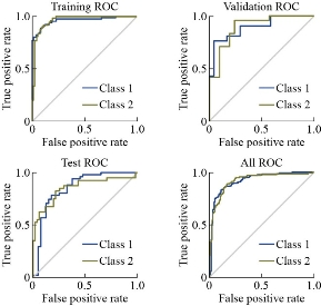
Fig. 7 Reciever operating characterstics curve using artificial neural network with multi-layer for Heart Disease Prediction
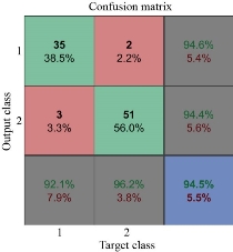
Fig. 8 Confusion matrices using deep sparse autoencoder based neural network for heart disease prediction.
Conclusions
In this work, we have developed a deep learning model for the prediction of heart disease. The performance of the deep learning can farther be enhanced by adding some more number of observations. As the dataset increases, the ability of the deep learning to learn the hidden features from the input will be improved. This study also indicates that the deep learning approach is more effective as compared to the traditional methods used for the diagnosis of heart disease. However, deep leaning has provided an exciting platform for fatal diseases risk assessment and opportunity to achieve higher accuracy in the medical field. The use of deep learning may assist the physician and doctors in a great deal and it may drive towards the personalized medicine.
Table 3Comparativeness of the Proposed Model with DNN
|
Model name
|
Train + Validation to test ratio |
Sensitivity |
Specificity |
Selectivity |
Detection rate |
F-measure |
Accuracy |
|
Yazdani, Armin, et al. [33]
|
- |
- |
- |
- |
- |
- |
96% Confidence score |
|
Almustafa, et al., [34]
|
- |
- |
- |
- |
- |
- |
93.8357% Decision Tree |
|
Shah, Devansh, et al., [35]
|
- |
- |
- |
- |
- |
- |
88.5% NBC 90.789% K-NN 80.263% Decision Tree 86.84% Random Forest
|
|
Jindal, Harshit, et al [36] |
- |
- |
- |
- |
- |
- |
87.5% Logistic Regression
88.5% KNN
|
|
ANN with 1 layer
|
70 – 30% |
80.9% |
61.2% |
64.1% |
71.05% |
71.5% |
77.6% |
|
ANN with 2 layers
|
70 – 30% |
72.3% |
86.3% |
85% |
79.3% |
78.13% |
85.5% |
|
Deep Neural Network
|
70 – 30% |
94.4% |
92.1% |
98.06% |
93.25% |
96.19% |
94.5% |
This work is supported by the National Key Basic Research Program (973 Project) (No.2017FYA 0205301), National Natural Scientific Fund (No. 81225010, 81327002, and 31100717), 863 Project of China (2014AA020700), Shanghai Science and Technology Fund (No. 13NM1401500 and 15DZ2252000).
Daxiang Cui conceived and designed the research project. Muhammad Aqeel Aslam, Muhammad Asif Munir, Rauf Ahmad, Muhammad Samiullah, Nasir Mahmood Hassan performed the experiments, data acquisition and finished the cardiovascular disease classification using deep neural networks. Daxiang Cui discussed the data analysis strategy, all the authors contributed to the data analysis and the writing of this manuscript, and all authors reviewed the manuscript and given approval to the final version of the manuscript.
The authors declare they have no conflict of interest, computing interest, financial or otherwise.
This research paper does not contain any studies with human participants or animals performed by any of the authors.
No human or animal is involved in this study.
[1].Benjamin, Emelia J et al. “Heart Disease and Stroke Statistics-2017 Update: A Report From the American Heart Association.” Circulation, vol. 135, no. 10, 2017.
[2].Park, Hyun Woo, et al. "A hybrid feature selection method to classification and its application in hypertension diagnosis." International Conference on Information Technology in Bio-and Medical Informatics. Springer, 11 – 19, Cham, 2017.
[3].Park, Hyun Woo, et al. "Risk factors rule mining in hypertension: Korean national health and nutrient examinations survey 2007–2014." 2016 IEEE Conference on Computational Intelligence in Bioinformatics and Computational Biology (CIBCB). IEEE, 1-4, 2016.
[4].World Health Organization (WHO). https://www.who.int/news-room/fact-sheets/detail/cardiovascular-diseases-(cvds). Accessed 01 August 2020
[5].Raza, Ali, et al. "Heartbeat sound signal classification using deep learning." Sensors, vol. 19, no. 21, 2019.
[6].Hanna, Ibrahim R., and Mark E. Silverman. "A history of cardiac auscultation and some of its contributors." The American journal of cardiology, vol. 90, no. 3, pp. 259-267, 2002.
[7].Chirakanphaisarn, Neramitr, ThadsaneeThongkanluang, and YuwathidaChiwpreechar. "Heart rate measurement and electrical pulse signal analysis for subjects span of 20–80 years." Journal of Electrical Systems and Information Technology, vol. 5, no.1, pp. 112-120, 2018.
[8].Jiang, Zhongwei, and Samjin Choi. "A cardiac sound characteristic waveform method for in-home heart disorder monitoring with electric stethoscope." Expert Systems with Applications, vol. 31, no..2, pp. 286-298, 2006.
[9].National Heart, Lung, and Blood Institute. https://www.nhlbi.nih.gov/health-topics/ coronary-heart-disease. Accessed 15August 2020
[10].Nucleus Medical Media. http://www.nucleushealth.com/Accessed 01 Oct 2019
[11].Hausmann, Harald, et al. "Decision-making in end-stage coronary artery disease: revascularization or heart transplantation." The Annals of thoracic surgery, vol. 64, no..5, pp. 1296-1302, 1997.
[12].Diamond, George A., and James S. Forrester. "Analysis of probability as an aid in the clinical diagnosis of coronary-artery disease." New England Journal of Medicine, vol. 300, no. 24, pp. 1350-1358, 1979.
[13].Zafar, Kashan, et al. "Skin lesion segmentation from dermoscopic images using convolutional neural network." Sensors, vol. 20, no..6, pp.1601-1614, 2020.
[14].Kim, Hyeongsoo, et al. "A data mining approach for cardiovascular disease diagnosis using heart rate variability and images of carotid arteries." Symmetry, vol. 8, no. 6, p.47, 2016
[15].Kim, Jaekwon, Jongsik Lee, and Youngho Lee. "Data-mining-based coronary heart disease risk prediction model using fuzzy logic and decision tree." Healthcare Informatics Research, vol. 21, no. 3, pp. 167-174, 2015.
[16].Kim, Jae Kwon, and Sanggil Kang. "Neural network-based coronary heart disease risk prediction using feature correlation analysis." Journal of Healthcare Engineering, pp. 1-13, 2017.
[17].Olaniyi, Ebenezer Obaloluwa, OyebadeKayodeOyedotun, and Khashman Adnan. "Heart diseases diagnosis using neural networks arbitration." International Journal of Intelligent Systems and Applications, vol. 7, no.12, p. 72, 2015.
[18]. Haq, Amin Ul, et al. "A hybrid intelligent system framework for the prediction of heart disease using machine learning algorithms." Mobile Information Systems, pp. 1-21, 2018. [19]. Shouman, Mai, Tim Turner, and Rob Stocker. "Using decision tree for diagnosing heart disease patients." Proceedings of the Ninth Australasian Data Mining Conference-Volume 121. 2011.
[20].Deekshatulu, B. L., and Priti Chandra. "Classification of heart disease using k-nearest neighbor and genetic algorithm." Procedia Technology, vol. 10, pp. 85-94, 2013.
[21].Ghumbre SU, Ghatol AA., Heart Disease Diagnosis Using Machine Learning Algorithm. In 2012 Proceedings of the International Conference on Information Systems Design and Intelligent Applications 2012, pp. 217-225, 2012.
[22].Sakr, Sherif, et al. "Comparison of machine learning techniques to predict all-cause mortality using fitness data: The Henry ford exercise testing (FIT) project." BMC medical informatics and decision making, vol. 17, no.1, p.174, 2017.
[23].Kourou, Konstantina, et al. "Machine learning applications in cancer prognosis and prediction." Computational and Structural Biotechnology Journal, vol. 13, pp. 8-17. 2015.
[24].Cifuentes-Alcobendas, Gabriel, and Manuel Domínguez-Rodrigo. "Deep learning and taphonomy: high accuracy in the classification of cut marks made on fleshed and defleshed bones using convolutional neural networks." Scientific Reports, vol. 9, no.1, pp.1-12, 2019
[25].Aslam, Muhammad Aqeel, et al. "Breath analysis based early gastric cancer classification from deep stacked sparse autoencoder neural network." Scientific Report, vol. 11, no. .1, pp:1-12, 2021.
[26]Hasan, Tabreer T., Manal H. Jasim, and Ivan A. Hashim. "FPGA Design and Hardware Implementation of Heart Disease Diagnosis System Based on NVG-RAM Classifier." 2018 Third Scientific Conference of Electrical Engineering (SCEE). IEEE, 2018.
[27].Muhammad, Yar, et al. "Early and accurate detection and diagnosis of heart disease using intelligent computational model." Scientific Reports, Vol. 10, no.1, pp. 1-7, 2020..
[28].Abdullah AS, Rajalaxmi R., A data mining model for predicting the coronary heart disease using random forest classifier. In 2012 International Conference in Recent Trends in Computational Methods, Communication and Controls, 22-25, 2012.
[29].Srinivas, K., B. Kavihta Rani, and A. Govrdhan. "Applications of data mining techniques in healthcare and prediction of heart attacks." International Journal on Computer Science and Engineering (IJCSE), vol. 2, no. 2, pp. 250-255, 2010.
[30].Nahar, Jesmin, et al. "Computational intelligence for heart disease diagnosis: A medical knowledge driven approach." Expert Systems with Applications, vol. 40, no.1, pp. 96-104, 2013.
[31].Chaurasia, Vikas, and Saurabh Pal. "Early prediction of heart diseases using data mining techniques." Caribbean Journal of Science and Technology, vol. 1, pp. 208-217, 2013.
[32]Das, Resul, Ibrahim Turkoglu, and Abdulkadir Sengur. "Effective diagnosis of heart disease through neural networks ensembles." Expert Systems with Applications, vol. 36, no.4, pp. 7675-7680, 2009.
[33].Yazdani, Armin, et al. "A novel approach for heart disease prediction using strength scores with significant predictors." BMC Medical Informatics and Decision Making, vol. 21, no.1, pp. 1-16, 2021.
[34].Almustafa, Khaled Mohamad. "Prediction of heart disease and classifiers’ sensitivity analysis." BMC bioinformatics, vol. 21, no.1, pp. 1-18, 2020.
[35].Shah, Devansh, Samir Patel, and Santosh Kumar Bharti. "Heart disease prediction using machine learning techniques." SN Computer Science, vol. 1, no.6, pp. 1-6, 2020.
[36].Jindal, Harshit, et al. "Heart disease prediction using machine learning algorithms." IOP Conference Series: Materials Science and Engineering. vol. 1022. no. 1. IOP Publishing, 2021.
[37].Bache, K., Lichman, M.: UCI Machine Learning Repository Irvine. University of California, School of Information and Computer Science, Oakland (2013).
[38].Kadam, VJ, Jadhav SM., Feature Ensemble Learning Based on Sparse Autoencoders for Diagnosis of Parkinson’s Disease. In 2019 Computing, Communication and Signal Processing. Advances in Intelligent Systems and Computing, pp. 567-581, 2019.
[39].Hinton, Geoffrey E., and Ruslan R. Salakhutdinov. "Reducing the dimensionality of data with neural networks." Science, vol. 313, no.5786, pp. 504-507, 2006.
[40].Bengio, Yoshua, and Yann LeCun. "Scaling learning algorithms towards AI." Large-scale Kernel Machines, vol. 34, no.5m pp. 1-41, 2007.
[41].Ranzato, Marc'Aurelio, et al. "Efficient learning of sparse representations with an energy-based model." Advances in Neural Information Processing Systems, vol. 19, pp. 1137-1144, 2006.
[42].Baldi P., Autoencoders, unsupervised learning, and deep architectures. In Proceedings of ICML Workshop on Unsupervised and Transfer Learning, pp. 37-50. 2012.
[43].Kittler, Josef, et al. "On combining classifiers." IEEE Transactions on Pattern Analysis and Machine Intelligence, vol. 20, no.3, pp. 226-239, 1998.
Copyright© Muhammad Aqeel Aslam, Muhammad Asif Munir, Rauf Ahmad, Muhammad Samiullah, Nasir Mahmood Hassan, Shahzadi Mahnoor, and Daxiang Cui. This is an open-access article distributed under the terms of the Creative Commons Attribution License, which permits unrestricted use, distribution, and reproduction in any medium, provided the original author and source are credited.

|
Nano Biomedicine and Engineering. Copyright © Shanghai Jiao Tong University Press |
 |