
Article
Synthesis of Ru(bpy)3-doped Silica Nanoparticle and Its Application in Fluorescent Immunoassay
Dongguang Yin *,Binhu Liu, Li Zhang,Chunjuan Xie, Le Zhang
College of Environmental and Chemical Engineering, Shanghai University, Shanghai
* Corresponding author. E-mail: ydg@shu.edu.cn
Citation: D. Yin et al., Study on Preparation of Ru(bpy)3-Doped Silica Nanoparticles and Their Application in Fluorescent Immunoassay. Nano Biomed Eng. 2010, 2(2): 117-120.
DOI: 10.5101/nbe.v2i2.p117-120
Abstract
A novel type of amino functionalized core-shell Ru(bpy)3-doped silica nanoparticles was synthesized using a simple and effective approach of reverse microemulsion. The nanoparticles were characterized by transmission electron microscope, fluorescence spectra, UV-Vis spectroscopy and tests of photostability and dye molecular leakage. It was found that the nanoparticles exhibit excellent fluorescent properties such as extremely bright, highly photostable and chemical stable. Furthermore, the nanoparticles utilized as a fluorescent marker applying in fluorescent immunoassay of mouse IgG were studied and desired results were obtained.
Keywords: Ru(bpy)3-doped; Silica nanoparticles ; Reverse microemulsion; Immunoassay
Currently, nanoparticles-based techniques shows great promise in applications of bioassay and biomedical [1-6]. Dye-doped silica nanoparticles have many unique properties such as highly fluorescent intensity, highly photostability, and excellent biological compati-bility[7,8]. Caused on the fact of these, preparation of new silica nanoparticles internally doped with organic and inorganic fluorophores and their application in bio-logical detection and immunoassay were widely investigated and effectively approached. In this study, we synthesized dye-doped nanoparticles using a water-in-oil microemulsion method by doping the fluorescent dye of tris-(2’2-bipyridyl) dichlororuthenium (II) hexahydrate (Ru(bpy)3) inside silica material. Approach of water-in-oil microemulsion has advantages of no requirement of extreme condition such as temperature and pressure. In addition, size and shape of the nanoparticles can be simply controlled by varying microemulsion parameters. A silane reagent of 3-aminopropyl-trimethoxysilane (APTMS) was first time used to prepare the nanoparticles. It directly introduced primary amino groups to surface of the nanoparticles by copolymerization reaction with tetraethyl orthosilicate (TEOS) [9]. After conjugating with streptavidin, immunoassay experiments using the nanoparticles as fluorescent probe were performed.
2. Materials and Methods
2.1 Materials and instruments
Ru(bpy)3, TritonX-100, mouse IgG, goat antimouse IgG, BSA, strepavidin (SA) were purchased from sig-ma. TEOS, APTMS, n-hexanol, cyclohexane and ammonium hydroxide, (28-30 wt %) were purchased from Shanghai Chemical Reagent Co. Ltd. (Shanghai, China). Ultrapure water (18 MΩ) was used for the preparation of all aqueous solutions. Unless otherwise stated, all chemicals and reagents used in this study were analytical grade quality. a Thermoflash multimode microplate reader with 452nm of excitation wavelength, 608nm of emission wavelength. Hitachi 800 transmission electron microscope(Japan) was used for measuring shape and size of nanoparticles. UV-Vis absorption spectra were recorded on a Hitachi U-3010 UV-Vis spectro-photometer (Japan). Fluorescence spectra were recorded on a Hitachi F-7000 spectrofluorometer (Japan). Fluorescence immunoassay was carried out with Corning Fluoro Nunc 96-well microtiter plate as the solid-phase carrier and measured on a Thermo flash multimode microplate reader with 452nm of excitation wavelength, 608 nm of emission wavelength.
2.2 Preparation of Ru(bpy)3-doped nanoparticles
Nanoparticles were synthesized by using a microemulsion method as Santra et al described [10] previously. Firstly, the microemulsion solution was pre-pared by mixing cyclohexane (7.5mL), n-hexanol (1.8mL), Triton X-100 (1.77μL), and water (400ul) with vigorous stirring. After mixing for 20 min, 0.5mg Ru(bpy)3 dye in water(0.1mL), 100μL TEOS and 5μL APTMS were added to the mixture. After stirring for 20 min, 60μL of NH4OH was added to initiate the polymerization reaction. The reaction was allowed to continue for 24h. When the polymerization was complete, an equal volume of acetone was added, and the mixture was vortexed to break the microemulsion state. The solidified silica nanoparticles were collected by centrifugation at 12000r/min for 10 min and ultrasonically washed with water and 95% ethanol two times to re-move residual surfactant and dye molecules, respectively. After air drying, desirable products of the nanoparticles were obtained.
2.3 Photo bleaching experiments and dye leaking experiments
To evaluate photostability of the nanoparticles, pho-tobleaching experiments of the nanoparticles and pure Ru(bpy)3 were performed in aqueous solution using a 150 W xenon lamp as an excitation source. The nano-particles were continuously exposed at 452 nm of max-imum excitation wavelength. Fluorescent intensities of the nanoparticles were recorded at every 15min interval for a period of 1.5 h on a spectrofluorometer with 591 nm of emission wavelength. To perform dyeleaking experiments, 1mg of the na-noparticles was dissolved in 10mL water and the mix-ture was ultrasonic dispersed continuously. At every 1h, 1mL of the suspending solution was taken out and cen-trifugated. After centrifugal separation, the precipitate was redissolved in 1ml water, ultrasonic dispersed, and then emission spectra were recorded. According to varies of the fluorescent intensity with time, dyeleaking of the nanoparticles in aqueous solution was evaluated.
2.4 Preparation of nanoparticles-labeled SA
To conjugate with SA [11], the nanoparticles were coated with BSA first. 1.0mg nanoparticles ultrasonic dispersed in 1.0mL of 0.1M phosphate buffer (pH 7.0) was mixed with 4.0mg of BSA and 0.3 mL of 1% (v/v) glutaraldehyde, stirred for 24 h at 4℃. After centrifuging and washing with the phosphate buffer two times, the BSA coated nanoparticles were suspended in 1.0mL of the phosphate buffer again, then 200 μg of SA and 0.2mL of 1% glutaraldehyde were added. After stirring at 4℃ for 24h, 2mg of NaBH4 was added, the reaction was allowed to continue for 2 h. After being centrifuged and washed with the phosphate buffer and water, the nanoparticle-labeled SA was further purified by gel filtration chromatography on a Sephadex G-50 column, eluting with mobile phase of 0.05M NH4HCO3 (pH 8.0). The fractions containing the nanoparticles-labeled SA were collected and stored at 4 ℃ after diluting with 0.1M phosphate buffer (pH 7.4)containing 0.1% BSA, 0.05% NaN3 and 0.9 % NaCl.
2.5 Coating biotinylated goat anti-mouseIgG to mi-crowell and fluorescent immunoassay
To coat biotinylated goat antimouse IgG to microwell, 200μL 10μg/mL of biotinylated goat anti-mouse IgG (in 0.5 M NaHCO3, pH 9.6) was added to each well of 96-microwells plates (black) and incubated for 24 h at 4 ℃. After twice washing with 0.05M phosphate buffer (pH 7.4, containing 0.9 % NaCl and 0.05 % Tween-20) three times, the microwells were blocked by incubation with 2 % BSA (w/v) for 2 h at room temperature. To perform fluorescent immunoassay, a serial dilution of nanoparticles-labeled SA in 0.1 M phosphate buffer (pH 7.0, containing 0.9 % NaCl and 0.1 % BSA) was added to each well and let the plate incubated for 3h at 37 ℃. After excess nanoparticles-labeled SA was removed thoroughly by washing four times in 0.05M phosphate buffer (pH 7.4, containing 0.9 % NaCl and 0.05 % Tween-20) with vigorous shaking, fluorescence intensities were measured in a THERMO Multiskan Ascent.
3. Results and Discussion
3.1 Characterization of the nanoparticles
TEM imaging of the nanoparticles are shown in Fig.2. Results show that the nanoparticles are spherical, monodisperse, and uniform in shape and size with av-erage diameter of 70±5nm. Black dots embedded inside the silica network can be observed due to the presence of heavy metal atom in the dye [12]. Absorption and fluorescence emission spectra of Ru(bpy)3 dye and the Ru(bpy)3-doped nanoparticles are shown in Fig.2A and Fig.2B. The absorption spectra of the pure Ru(bpy)3 dye and the nanoparticles display a same profile in aqueous solution. Their emission spectra also show a same profile but maximum emission wavelength of the nanoparticles shift 8nm, toward the shorter wavelength.

Figure1. Transmission electron micrograph images of Ru(bpy)3-doped silica nanoparticles
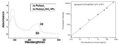
Figure 2. Absorption spectrum(A) and emission spectrum (B) of the Ru(bpy)3-doped nanoparticle and pure Ru(bpy)3

Figure 3. Results of confirmation of -NH2 groups on surface of the nanoparticles
3.2 Confirmation of amino groups on surface of the nanoparticles
When ninhydrin reacts with amino compound, a blue-violet compound is produced, which has a absorbance peak at about 570 nm [13]. Based on this fact, confirmation of amino groups on surface of the nanoparticles was investigated. Pure APTMS and two types of Ru(bpy)3-doped nanoparticles were compared. One type of the nanoparticles was prepared through copolymerization of APTMS and TEOS, another type of the nanoparticles was prepared through polymerization of TEOS but without APTMS. As shown in Fig.4, pure APTMS and the former nanoparticles all have maximal absorbance peaks at 563nm, whereas the nanoparticles prepared without APTMS have non- absorbance peak at 563nm. The results demonstrated that amino groups had been introduced to the surface of the nanoparticles directly during the preparation process. Since the nano-particles have active amino groups on their surface, they can be directly used to conjugate biological molecules without complicated surface modification.
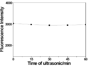
Figure 4. Results of photo stability experiments of the nanoparticles
3.3 Photostability and dyeleaking of the nanoparticles
As shown in Figure 4, the fluorescent intensity of pure Ru(bpy)3 was decreased approximately 57% after 1.5h of continuous excitation, whereas the fluorescent intensity of the nanoparticles was only decreased 10%. The high photostability of the nanoparticles is due to the fact that the Ru(bpy)3 dye in the nanoparticles is coated surroundingly by silica, which isolates the dye molecules from the outside environment such as sol-vent molecules, oxygen and free radicals caused by light exposure. Therefore, effectively protects the dye molecules from photodecomposition. As shown in Figure 5, the fluorescent intensity of the nanoparticles was only decreased approximately 0.2% after continuously ultrasonic 60min in aqueous solution. The results indicate the nanoparticles are stable in aqueous solution.
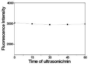
Figure 5. Results of dyeleaking experiments of the nanoparticles
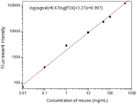
Figure 6. Results of fluorescent immunoassay using the nanoparticles as fluorescentprobes.
3.4 Application of the nanoparticles to fluorescent immunoassay
This assay system is identical to that of ELISA except that fluorescent nanoparticles takes the place of enzyme. After the coated plates were blocked with BSA, the SA-nanoparticles as a probe was added to the 96-well microtiter plate. After unbound SA-nanoparticles were washed away, the fluorescence in-tensities were measured. Results shown in Fig. 6 indicate a highly specific binding of the SA-nanoparticles to the biotinylated goat antimouse IgG. The relation-ship between the fluorescence intensities and the SA-nanoparticles concentrations was linear in the experimental range. These results demonstrate the potential to apply this newly developed fluorescent nanoparticles in various sensitive immunoassay.
Acknowledgements
Financial support from the Shanghai Pujing Program, Shanghai Nano Program (No. 0752nm024), Nation 973(2010CB933901), and Shanghai Leading Academic Discipline Project (No. S30109) is gratefully acknowledged.
References
Received 26 May,2010;accepted 10 June, 2010; published online 26 June, 2010.
Copyright: (C) 2010 D. Yin et al. This is an open access article distributed under the terms of the Creative Commons Attribution License, which permits unrestricted use, distribution, and reproduction in any medium, provided the original author and source are credited.

|
Nano Biomedicine and Engineering. Copyright © Shanghai Jiao Tong University Press |
 |